In the final part of our series on bi-specific antibodies, we’ll discuss some of the assays that are used to test the specificity, functionality, and safety of bi-specific antibodies.
How are bi-specific antibodies tested for antigen specificity?
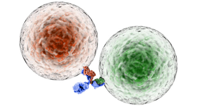
As described in our previous blogs, bi-specific antibodies have the capability to recognize two different antigens due to the presence of two different antigen recognition domains. To ensure that each arm has specificity for the correct antigen, binding assays are performed with a cell line that expresses the antigen recognized by one arm of the bi-specific antibody. Typically, flow cytometry is used as a readout to show the bi-specific antibody is binding the antigen-expressing line in a titratable manner. A further requirement in these assays is the addition of a competitive antibody that also has specificity for the antigen, which should block binding of the bi-specific antibody. Together, these binding assays serve to test the specificity of the bi-specific antibodies for their cognate antigens. Please see iQ’s web page for more information regarding our binding assay bioservices.
What are some assays to test for functionality of bi-specific antibodies?
In many cases, the goal of a bi-specific antibody is to recruit an immune cell to the target cell to lyse it. Therefore, cytotoxicity assays are performed to demonstrate killing of targets by immune cells. There are many readouts for cytotoxicity assays, including the Calcein release assay. In this assay, target cells are loaded with Calcein-AM, a molecule which becomes fluorescent once inside the cell, that is released into the supernatant upon cell death. The supernatant is then collected and assessed for fluorescence, which is directly proportional to cytotoxicity. In a typical experiment, immune cells are cultured with Calcein-loaded target cells in the presence of the bi-specific antibody. After the culture period, the supernatant is collected and analyzed for fluorescence. With this assay, the entire plate can be read at once providing a high throughput method to measure cytotoxicity.
Another fairly high throughput assay for cytotoxicity is a flow cytometric-based method that measures cell death. Here, immune cells are initially loaded with a fluorescent dye, such as CFSE, to distinguish them from targets during flow acquisition. These labeled immune cells are cultured with targets in the presence of the bi-specific antibody, and then stained with propidium iodide or annexin V, which label dead cells. The samples are analyzed by flow cytometry for cell death based on propidium iodidie or annexin V incorporation.
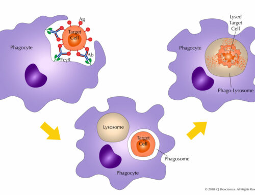
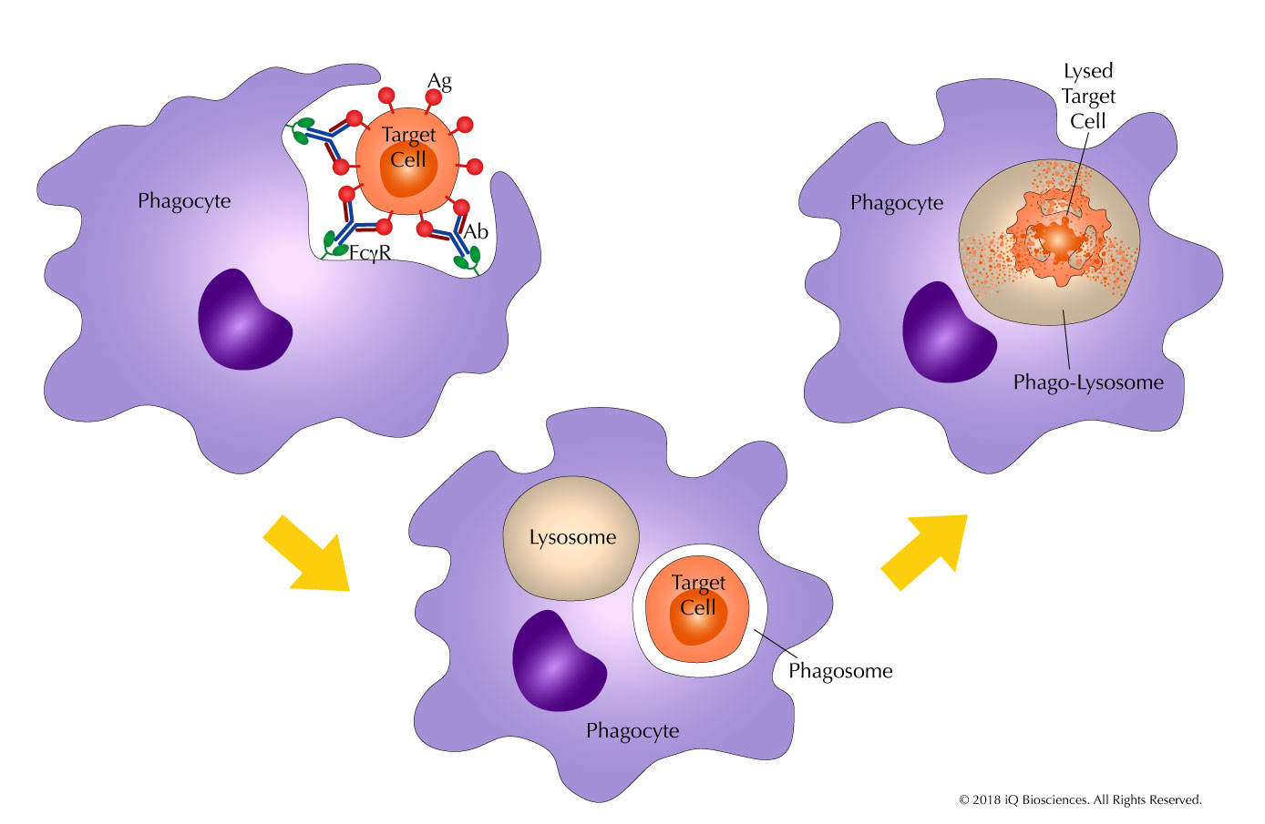
Additionally, bi-specific antibodies that retain the Fc portion of an antibody may also have Fc-mediated effector functions, such as antibody-dependent cellular cytotoxicity (ADCC). In contrast to the cytotoxicity assays described above, cytotoxicity cannot be used as a readout since cytotoxicity may be driven in a non-Fc receptor mediated manner. Therefore, an artificial cell line that only expresses the Fc receptor without any other activating receptors is used. In addition, this cell line must also be able to report Fc receptor-mediated activation. Fortunately, there are proprietary cell lines that express the Fc receptor and when activated through it, express luciferase. In these assays, the reporter cell lines are cultured with targets and the bi-specific antibody. After the culture period, luciferase expression, which is directly proportional to Fc receptor activation, is measured on a luminometer. Please see our blog and website for more information regarding iQ’s ADCC assay bioservice.
Are there other assays beside cytotoxicity to assess function?
A bi-specific antibody can also serve other purposes in addition to recruitment of an immune cell to its target. In some instances where one of the arms recognizes CD3, the bi-specific antibody can also help activate T cells to produce cytokines. Thus, another method to assess bi-specific antibody mediated function is to assay for cytokines, which can be performed using a cytokine release assay (CRA). In these assays, T cells are incubated with the bi-specific antibody and then the supernatant is collected after the culture period. Using a cytokine bead array kit, the supernatant can be interrogated for multiple secreted factors simultaneously. The cytokines or chemokines observed in the supernatant can lend insight into the type of response that is produced, such as an inflammatory one.
The CRA can also be used to assess safety of the bi-specific antibody in cases where an inflammatory response is not preferred. Like with other therapeutic molecules, there is the possibility of eliciting a massive release of cytokine, which could lead to Cytokine Release Syndrome, a potentially life-threatening event, after administration. In some trials, mild cases of CRA are observed after infusion. The assay is the same as described above except the hope is the absence of cytokine release when the bi-specific antibody is cultured with T cells. Please see our blog and website for more information regarding iQ’s CRA bioservice.
Together, these assays can assess the specificity, function, and safety profile of bi-specific antibodies. Choosing an immunology-based CRO, such as iQ Biosciences, who has the experience and resources to perform these functional experiments is important. The selection of an experienced and knowledgeable CRO will help generate data for the development of these molecules that are beginning to show much promise.
