In an antibody-drug conjugate (ADC) experiment, the choice of isotype control shapes how clearly target-specific activity can be separated from background. An isotype control accounts for effects that are not specific to the target, which makes choosing the right one an important part of experimental design.
Our previous blog post covered why a matched ADC isotype control matters and which properties should align with the molecule under study. This article builds on that foundation with a decision that gets less attention. When should an isotype control use a wild-type IgG1 backbone, and when should it be Fc-silent? The answer depends on the assay, and it also connects to a broader question in ADC development, whether Fc effector function helps or hurts a given therapeutic.
What an isotype control accounts for
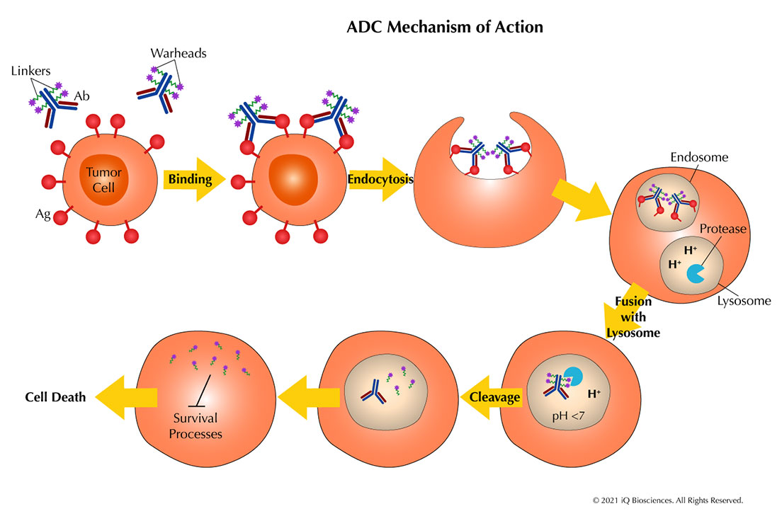
An ADC combines an antibody, a linker, and a cytotoxic payload. Each of these components can contribute to activity in an assay, and not all of that activity is target-specific. The antibody can bind non-specifically or engage Fc receptors, and the payload brings its own background cytotoxicity independent of the target. An isotype control is a non-targeting antibody, or ADC, that carries the same non-specific properties as the molecule under study without binding the intended antigen.
Used this way, the isotype control establishes a baseline. Signal above that baseline can be attributed to target engagement, while signal shared with the control reflects non-specific antibody interactions, Fc receptor binding, or payload-driven background (Figure 1). The closer the control matches the ADC in its non-target properties, the more informative that baseline becomes.

Figure 1. Antibody with payload and linker schematic
Matching the control to the ADC
Three properties are generally matched between an isotype control ADC and the molecule under study: the payload, the drug-to-antibody ratio (DAR), and the conjugation chemistry. The payload contributes much of an ADC’s background cytotoxicity, so matching it keeps payload-driven effects in the baseline. Matching the DAR, which influences potency and the conjugate’s physical behavior, gives a comparable baseline. Conjugation chemistry matters too, since the way a payload is attached affects the properties of the conjugate. When these align, differences between the ADC and its control more likely reflect target-mediated activity (Figure 2).

Figure 2. Payload, DAR, Conjugation Chemistry & Linker
With payload, DAR, and conjugation chemistry matched, the antibody backbone is one important decision that remains. This is where the choice depends most directly on the biology being studied and on the design of the therapeutic ADC itself.
Wild-type and Fc-silent backbones
Isotype control ADCs are available on both wild-type and Fc-silent human IgG1 backbones. The two are identical in the Fab region and differ only in the Fc, which determines how they interact with Fc-gamma receptors (FcγRs) and the complement protein C1q (Figure 3).

Figure 3. IgG1 Wildtype versus Fc- silent (LALAPG) IgG1 backbones
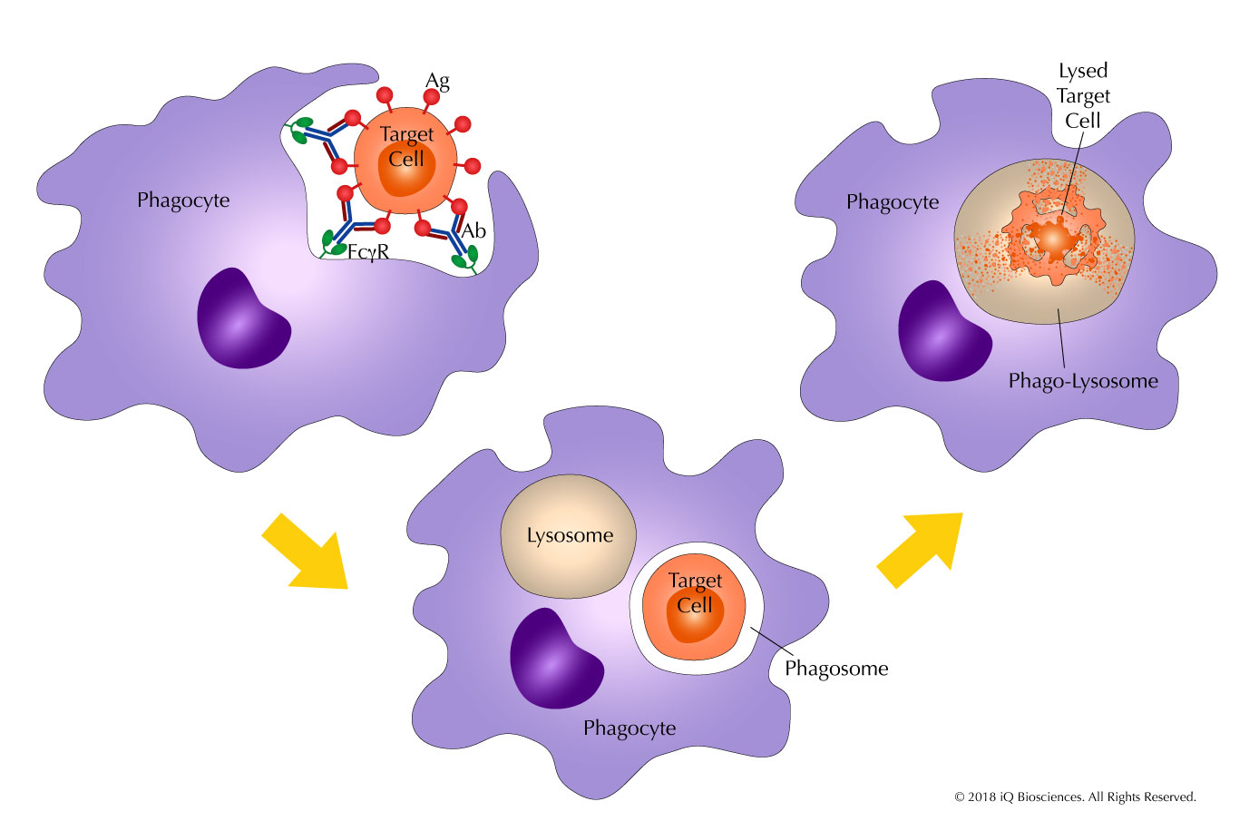
A wild-type IgG1 backbone retains normal Fc function. It engages activating Fc-gamma receptors and binds C1q, and so it can participate in antibody-dependent cellular cytotoxicity (ADCC), antibody-dependent cellular phagocytosis (ADCP), and complement-dependent cytotoxicity (CDC), consistent with native IgG1 behavior.
An Fc-silent backbone is one that has been engineered to reduce or abolish Fc-mediated effector function. Several approaches exist, including aglycosylation (e.g., N297A/G), IgG2/IgG4 subclass frameworks, and various point-mutation combinations in the lower hinge and CH2 domain [3]. Among the most widely used is the LALAPG set of mutations — three substitutions (L234A, L235A, and P329G) in the Fc region [1] [2]. These mutations nearly eliminate binding to activating Fc-gamma receptors and C1q, so the antibody does not appreciably support ADCC, ADCP, or CDC [2] [3]. Binding to the neonatal Fc receptor (FcRn) is retained, which preserves the long circulating half-life associated with IgG, and thermal stability is largely retained, though modest reductions relative to wild-type IgG1 have been reported [1] [2] [3]. The result is a backbone that maintains the antibody’s structure and pharmacokinetic profile while removing its effector engagement.
It is worth noting why LALAPG has become a preferred Fc-silencing approach for many applications. The earlier LALA variant (L234A/L235A alone) reduces but does not fully silence Fc effector function. Hale (2024) showed that LALA retains approximately 78% of wild-type FcγRI activity and up to 28% of FcγRIIIa activity in cell-based assays, meaning it is incompletely silenced [3]. The addition of P329G in the LALAPG combination reduces these residual interactions to near-undetectable levels in most assay formats [2] [3], which is why LALAPG is generally favored when more complete effector silencing is the goal.
Fc effector function in ADCs: a double-edged sword
The choice between a wild-type and Fc-silent backbone is not just an assay design question. It reflects a broader issue in ADC development: whether Fc effector function is an asset or a liability for a given therapeutic. Understanding both sides of this equation helps explain why the backbone of the isotype control should match the backbone of the ADC under study (Figure 4).

Figure 4. Schematic comparing dual role of Fc function in ADCs
When Fc function is a liability
A growing body of evidence indicates that FcγR-mediated uptake of ADCs by off-target immune cells is a significant driver of dose-limiting toxicities. Taylor and Lindorfer (2024), in a perspective published in Blood, argued that ADCs can be cleared by the same machinery the immune system uses for immune complexes — phagocytic and trogocytic uptake by cells the ADC was never meant to reach — and that this misrouting accounts for much of the observed toxicity [4].
Several specific toxicities have been linked to target-independent, Fc-mediated uptake of ADCs by off-target cells. The table below summarizes the proposed mechanisms and key supporting references.
Table 1. Reported off-target toxicities of ADCs and their proposed Fc- and update-related mechanisms.
| Toxicity | Proposed Mechanism | ADC Examples | Key References |
|---|---|---|---|
| Thrombocytopenia | FcγRIIa-mediated uptake by megakaryocytes | T-DM1, gemtuzumab ozogamicin | Uppal et al. (2015) [5] |
| Thrombocytopenia / Hepatotoxicity | Target-independent platelet sequestration and sinusoidal endothelial cell injury in NHP model | Non-targeting calicheamicin conjugate | Guffroy et al. (2017) [6] |
| Neutropenia | Differentiating neutrophils killed by cytotoxic payload via linker cleavage [7] or trogocytosis [4] (proposed) | Multiple ADCs | Zhao et al. (2017) [7]; Taylor & Lindorfer (2024) [4] |
| Hepatotoxicity / SOS | FcγR-mediated uptake by liver sinusoidal endothelial cells (proposed) | Gemtuzumab ozogamicin, inotuzumab ozogamicin | Taylor & Lindorfer (2024) [4] |
| ILD / Pneumonitis | FcγR-mediated uptake by alveolar macrophages | T-DM1, T-DXd | Nguyen et al. (2023) [8] |
Experimental work supports the idea that Fc-silencing can mitigate some of these effects. Aoyama et al. (2022) showed that aggregated ADCs are taken up by FcγR-expressing cells as immune complexes, and that silencing the Fc region suppressed this off-target cytotoxicity [9]. In a more recent example, Kim et al. (2026) demonstrated that a non-targeting ADC on a wild-type backbone killed FcγR-expressing cells at low nanomolar concentrations, while the same conjugate on an effector-silenced backbone showed no detectable killing [10]. These findings, together with the clinical toxicity data, have prompted interest in Fc-silent ADC backbones as a potential mitigation strategy. To date, however, Fc silencing of ADCs remains largely unexplored in the clinic, where approved ADCs are almost uniformly built on wild-type IgG1 [3].
When Fc function is desired
Fc effector function is not always a liability. For some ADCs, it is part of the therapeutic mechanism, and removing it could reduce efficacy.
Both trastuzumab emtansine (T-DM1) and trastuzumab deruxtecan (T-DXd) retain the Fc effector functions of their parent antibody, trastuzumab, and operate through multiple mechanisms of action — including HER2 signaling blockade, cytotoxic payload delivery, and Fc-mediated immune functions such as ADCC and ADCP [11] [12].
T-DXd illustrates this particularly well. Tsao et al. (2025) demonstrated that T-DXd engages FcγRs and promotes ADCP of HER2-positive breast cancer cells by macrophages [13]. In the same study, the DXd payload was shown to trigger immunogenic cell death in tumor cells, which in turn activated dendritic cells and macrophages and enhanced their ability to present tumor antigens to CD8+ T cells [13]. The result is a convergence of two distinct immune-activating mechanisms — one driven by the Fc region, the other by the payload — that together amplify the anti-tumor immune response. This represents a case where Fc function actively contributes to the therapeutic effect rather than merely being tolerated.
Nessler et al. (2018) framed the underlying question clearly: are ADCs best understood as “armed antibodies,” where immune engagement drives efficacy, or as “targeted small molecules,” where payload delivery dominates? [12]. The answer likely varies by ADC, and this is precisely why the Fc backbone choice matters, both in the design of the therapeutic and in the selection of its matched isotype control.
Matching the backbone to the assay
With this context in mind, the practical question for assay design becomes clearer. The backbone choice matters because Fc engagement can itself generate signal in a cell-based assay, independent of the payload. Many cell types, including immune effector cells such as NK cells, macrophages, and neutrophils, express Fc-gamma receptors. An antibody with a functional Fc engages those receptors, which can drive internalization, cytokine release, or cytotoxicity unrelated to the target.
In an assay that includes FcγR-expressing cells, a wild-type control brings this Fc-driven contribution into the baseline along with the payload-driven one. That suits studies where Fc effector activity is itself part of the biology being measured, for example when evaluating an ADC whose therapeutic mechanism includes ADCC or ADCP. To isolate payload-driven activity instead, an Fc-silent control removes the Fc contribution, leaving a baseline of payload-driven and non-specific background without FcγR signaling. This is why Fc-silent backbones are commonly used for cytotoxicity, internalization, and bystander-effect studies in systems that contain FcγR-expressing cells.
The guiding principle is straightforward: match the isotype control backbone to the therapeutic ADC. If the ADC under study uses a wild-type IgG1 backbone, because Fc effector function is part of its intended mechanism, the isotype control should also be wild-type. If the ADC uses an Fc-silent backbone, the control should be Fc-silent. In both cases, the control is still matched to the ADC on payload, DAR, and conjugation chemistry.
Looking ahead
The question of whether ADCs should retain or silence Fc effector function is an active area of investigation. As the field accumulates more clinical data on Fc-mediated toxicities and more preclinical evidence on the immune contributions to ADC efficacy, the design of next-generation ADCs will increasingly reflect deliberate choices about the Fc backbone. Those choices will, in turn, determine the appropriate isotype controls for the studies that support them.
Isotype control ADCs at iQ Biosciences
iQ Biosciences offers isotype control ADCs on both wild-type and Fc-silent (LALAPG) human IgG1 backbones, available in GGFG-DXd, SMCC-DM1, and vc-MMAE payload formats. Matched isotype controls can also be generated to correspond to a specific ADC’s payload, DAR, and conjugation chemistry, and each conjugate is characterized by SEC, HIC, and LC-MS as part of the standard data package.
The full set of isotype control ADCs is available here.
References
[1]: Schlothauer, T. et al. (2016). Novel human IgG1 and IgG4 Fc-engineered antibodies with completely abolished immune effector functions. Protein Engineering, Design and Selection, 29(10), 457–466. https://doi.org/10.1093/protein/gzw040
[2]: Lo, M. et al. (2017). Effector-attenuating substitutions that maintain antibody stability and reduce toxicity in mice. Journal of Biological Chemistry, 292(9), 3900–3908. https://doi.org/10.1074/jbc.M116.767749
[3]: Hale, G. (2024). Living in LALA land? Forty years of attenuating Fc effector functions. Immunological Reviews, 328(1), 422–437. https://doi.org/10.1111/imr.13379
[4]: Taylor, R.P. & Lindorfer, M.A. (2024). Antibody-drug conjugate adverse effects can be understood and addressed based on immune complex clearance mechanisms. Blood, 144(2), 137–144. https://doi.org/10.1182/blood.2024024442
[5]: Uppal, H. et al. (2015). Potential mechanisms for thrombocytopenia development with trastuzumab emtansine (T-DM1). Clinical Cancer Research, 21(1), 123–133. https://doi.org/10.1158/1078-0432.CCR-14-2093
[6]: Guffroy, M. et al. (2017). Liver microvascular injury and thrombocytopenia of antibody-calicheamicin conjugates in cynomolgus monkeys — mechanism and monitoring. Clinical Cancer Research, 23(7), 1760–1770. https://doi.org/10.1158/1078-0432.CCR-16-0939
[7]: Zhao, H. et al. (2017). A potential mechanism for ADC-induced neutropenia: role of neutrophils in their own demise. Molecular Cancer Therapeutics, 16(9), 1866–1876. https://doi.org/10.1158/1535-7163.MCT-17-0133
[8]: Nguyen, T.D., Bordeau, B.M. & Balthasar, J.P. (2023). Mechanisms of ADC toxicity and strategies to increase ADC tolerability. Cancers, 15(3), 713. https://doi.org/10.3390/cancers15030713
[9]: Aoyama, M. et al. (2022). Fcγ receptor-dependent internalization and off-target cytotoxicity of antibody-drug conjugate aggregates. Pharmaceutical Research, 39, 89–103. https://doi.org/10.1007/s11095-021-03158-x
[10]: Kim, S. et al. (2026). A stealth-body Fc engineering platform enables complete effector silencing in antibody and ADC therapeutics. Journal of Biological Engineering (in press). https://doi.org/10.1186/s13036-026-00735-9
[11]: Hunter, F.W. et al. (2020). Mechanisms of resistance to trastuzumab emtansine (T-DM1) in HER2-positive breast cancer. British Journal of Cancer, 122, 603–612. https://doi.org/10.1038/s41416-019-0635-y
[12]: Nessler, I., Khera, E. & Thurber, G.M. (2018). Quantitative pharmacology in antibody-drug conjugate development: armed antibodies or targeted small molecules? Oncoscience, 5(5–6), 161–163. https://doi.org/10.18632/oncoscience.435
[13]: Tsao, L.-C. et al. (2025). Effective extracellular payload release and immunomodulatory interactions govern the therapeutic effect of trastuzumab deruxtecan (T-DXd). Nature Communications, 16, 3167. https://doi.org/10.1038/s41467-025-58266-8