- CD3+ T cells expanded from cynomolgus macaque purified T cells
- Expanded T cells are validated for retention of functional activity such as proliferation, cytokine production, and cytolytic activity
- Applications include a wide variety of safety assessment and functional assays
- All samples are isolated from CITES-approved donors, making these cells an ideal product for international purchase
- Carefully cryopreserved to ensure high viability (> 90%) upon thawing
- All orders come with an iQ Certificate of Analysis
Expanded Cynomolgus Monkey CD3 T Cells
$820.00 – $1,450.00
Description
About the Cynomolgus Monkey (or Cynomolgus Macaque)
The cynomolgus macaque (“cyno”) is the most utilized non-human primate in biomedical research. According to some researchers, these monkeys are ideal models due to their genetic similarity to and recent evolutionary divergence from humans. They are employed in numerous biomedical research areas, such as immunology, neuroscience, oncology, diabetes, and pharmacology due to their metabolic and physiological similarity to humans. CD3+ T cells represent a relevant cell subset in the pathology of many common diseases, and cyno CD3+ T cells are a valuable resource to help model cellular responses to various treatments.
Cyno CD3+ T cell Potential Application Summary
- Flow cytometry-based binding studies for target binding and cross-reactivity
- Large-scale potency assays to compare a panel of test articles for the ability to elicit T cell responses
- Adoptive transfer into immunodeficient recipient mice to support the in vivo evaluation of activity with therapeutic test articles
Expansion of Cyno CD3+ T cells
These cyno CD3+ T cells are meticulously expanded over a multi-day period using a combination of cytokines, expansion medium, and high quality T cell activation beads. The expansion process takes place in a controlled environment to ensure optimal conditions for cell growth and retained function.
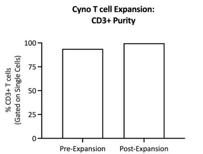
(a)
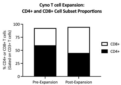
(b)
Figure 1: T cell phenotype is maintained post-expansion. (a) CD3+ purity tends to increase after expansion. (b) CD4+ T cell frequency slightly decreases after expansion with a concordant increase in CD8+ T cell frequency. Data shows one representative sample.
Validation of Expanded Cyno CD3+ T cells
Our expanded cyno CD3+ T cells have undergone rigorous testing to validate their exceptional quality and functional ability as reflected by their capacity for robust proliferation, target cell killing, and cytokine production, in vitro. These cells may be used for various applications, including mechanistic research, therapeutic development, and innovative cellular assays.
Proliferation
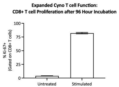
Figure 2: CD8+ T cells within expanded cyno CD3+ T cells proliferate in response to stimulation. Expanded cyno T cells were stimulated with activation beads for 96 hours. Flow cytometry was used to assess the frequency of Ki-67+ (a marker of proliferation) cells within CD8+ T cells. Data shows the average (mean) of technical triplicates with SEM error bars.
Cytokine Release
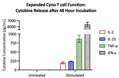
Figure 3: Expanded cyno CD3+ T cells secrete cytokines upon activation. Expanded cyno T cells were stimulated with activation beads for 48 hours. Cell-free supernatant was harvested and cytokine secretion was quantified via cytometric bead array. In response to stimulation, expanded T cells secrete IL-2, IL-10, TNF-α, and IFN-γ. Data shows the average (mean) of technical triplicates with SEM error bars.
T cell Dependent Cellular Cytotoxicity (TDCC)
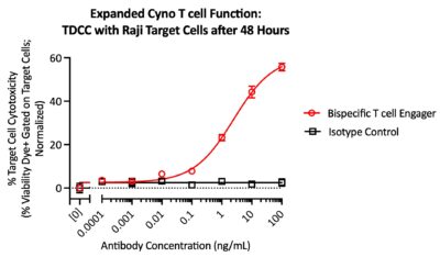
Figure 4: Expanded cyno CD3+ T cells exhibit cytotoxicity toward tumor target cells. Expanded cyno CD3+ T cells were co-cultured with fluorescently-labeled Raji cells and treated with either a cyno-reactive bispecific T cell engager antibody (CD3xCD20) or an isotype control. Raji target cell cytotoxicity was measured on a flow cytometer via viability dye incorporation. Data shows the average (mean) of technical triplicates, normalized with background subtraction, with SEM error bars.
Cryopreservation and storage
Expanded cyno CD3+ T cells are carefully cryopreserved using iQ Biosciences’ cryopreservation protocol that ensures high viability (> 90%) upon thaw.
Cells should be stored at < -120 °C once they are received, such as within a liquid nitrogen tank (vapor phase).
Additional information
| Available Size(s) | |
|---|---|
| Cell Type | |
| Format | |
| Species | |
| Tissue Type | |
| Viability | > 90% |
We are Committed to Ethical Practices
iQ Biosciences’ primary cell products from non-human primates are collected under approved IACUC protocol, which was developed in consultation with the Attending Veterinarian and is consistent with current veterinary standards. The collection facility is AAALACi and PHS accredited, and all animal housing, handling, and research protocols are consistent with standards set forth by the Animal Welfare Act, NIH's Policy on Humane Care and Use of Animals and the Guide for the Care and Use of Animals. All animal housing and handling is done in such a way to minimize stress and risk of injury to the animal.
Tailored Experiments
Your research is important to us. Our scientists will work through each step of the process with you, including assay design, data analysis, and recommendations for future studies.
Streamlined Process
Simplify your workflow. Bypass the middle-man: at iQ Biosciences, you'll get immediate access to our biospecimen inventory, saving you both valuable time and money.
Expertise
We're experts- so you don't have to be. Augmenting years of experience in immunology and working with immune assays, our scientists stay current with the latest publications and technology.
Exceptional Service
We're here to help. We know the challenges you're facing: whether it be through expedited service or our complimentary consulting services, our team is dedicated to helping you reach your goals.
For US customers, we ship via FedEx Overnight Shipping. Shipping charges will vary per shipping address (based on ZIP code) and are estimated to be $140.
For international (non-US) customers, we work closely with you and our couriers to ensure all necessary documentation is in place for international shipments to significantly reduce the chance of delays at Customs. For the export of non-human primate samples, this includes preparing CITES permits, as well as any other documentation as required by country. Please submit an inquiry to orders@iqbiosciences.com for your estimated time of delivery and shipping charges.
Austria
Hölzel Diagnostika Handels GmbH
Tel: +49 221 126 02 66
Email: info@hoelzel.de
Web: https://www.hoelzel-biotech.com/
Canada
Cedarlane
Tel: +1 (289) 288-0001
Toll Free (North America): +1 (800) 268-5058
Fax: +1 (289) 288-0020
Email: sales@cedarlanelabs.com
Web: https://www.cedarlanelabs.com
China
BIOHUB INTERNATIONAL TRADE CO., LTD.
上海起发实验试剂有限公司
Address: Chuansha Rd #6619, Pudong, Shanghai, Zipcode: 201200 P.R.China
Tel: 0086-021-50724187
Phone: +86-15921799099
Fax: 0086-021-50724961
Email: sale3@78bio.com
Web: www.qfbio.com
European Union
Caltag Medsystems Ltd.
Email: office@caltagmedsystems.co.uk
Web: https://www.caltagmedsystems.co.uk
tebu-bio
Web: https://www.tebu-bio.com
Or Find a local contact
Germany
Hölzel Diagnostika Handels GmbH
Tel: +49 221 126 02 66
Email: info@hoelzel.de
Web: https://www.hoelzel-biotech.com/
Zageno
Web: https://zageno.com/
Ireland
2B Scientific Ltd
Tel: +44(0) 1869 238033
Fax: +44(0) 1869 238034
Email: sales@2BScientific.com
Web: https://www.2bscientific.com
India
Cell & Gene BioSolutions Pvt. Ltd.
#478 C, SLV Complex, Raghavendra Swamy Mutt Road
Opp. Turahalli Water Tank, Turahalli, Subramanyapura Post
Uttarahalli Hobli, Bengaluru-560061, Karnataka, India
Phone: +91 97317 14670
Phone: +91 98809 25033
Email: info@cgbios.com
Web: www.cgbios.com
Japan
Cosmo Bio Co., Ltd.
Tel: +81 (03) 5632 9610
Fax: +81 (03) 5632 9619
Email: nsmail@cosmobio.co.jp
Web: https://www.cosmobio.co.jp
Qatar
Sedeer Medical Services and Trading LLC
Tel: +974 4434 9191
Email: info@sedeer.com
Web: https://sedeer.com/
Singapore
Omnicell Pte Ltd
Tel: +65 6747 0201
Email: enquiry@omnicell.com.sg
Web: https://omnicell.com.sg/</a
South Korea
BioClone
Tel: +82-2-2690-0058
Email: bioclone@bioclone.co.kr
Web: http://www.bioclone.co.kr
Switzerland
Hölzel Diagnostika Handels GmbH
Tel: +49 221 126 02 66
Email: info@hoelzel.de
Web: https://www.hoelzel-biotech.com/
Taiwan
Hycell International Co. Ltd.
Tel: +886-2-2877-1122
Fax: +886-2-2876-1520
Web: http://www.hycell.com.tw
United Kingdom
2B Scientific Ltd
Tel: +44(0) 1869 238033
Fax: +44(0) 1869 238034
Email: sales@2BScientific.com
Web: https://www.2bscientific.com
Caltag Medsystems Ltd.
Tel: +44 (0)1280 827460
Fax: +44 (0)1280 827466
Email: office@caltagmedsystems.co.uk
Web: https://www.caltagmedsystems.co.uk
tebu-bio
Tel: +44 (0)1733 421880
Fax: +44 (0)1733 421882
Email: uk@tebu-bio.com
Web: https://www.tebu-bio.com
Zageno
Web: https://zageno.com/
United States
Fisher Scientific
Tel: 1-800-766-7000
Web: https://www.fishersci.com
Quartzy
Web: https://www.quartzy.com
VWR International
Tel: 1-800-932-5000
Web: https://www.vwr.com
Zageno
Web: https://zageno.com/
Related products
-
Purified Cynomolgus Monkey CD14+ Cells
$880.00 – $1,695.00 -
Purified Rhesus Macaque Monkey CD4 T Cells
$1,840.00 – $3,070.00 -
Human Peripheral Blood Mononuclear Cells (PBMCs)
$330.00 – $440.00 -
Purified Rhesus Monkey CD14+ Cells
$700.00 – $1,345.00
