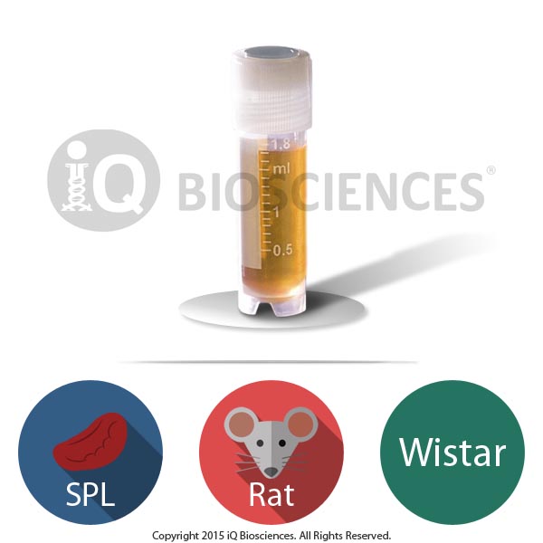
- CD3 Pan T cells purified from Sprague Dawley rat PBMCs
- Greater than 95% post-selection purity achieved via magnetic isolation
- Used for a wide variety of safety assessment and functional assays for research and nonclinical drug development
- Carefully cryopreserved to ensure high viability (> 70%) upon thawing
Purified SD Rat CD3 Pan T Cells
$410.00
 Powered by Bioz
Description
About the Sprague Dawley Rat
The Sprague Dawley (SD) rat is one of the two most widely-used outbred rat strains in biomedical research, including oncology, neurobiology, toxicology, pharmacology, aging, metabolism/nutrition, and neurobiology. Derived from the Wistar rat strain, the Sprague Dawley rat is also routinely employed in the field of immunology research as a model for autoimmune diseases and immunogenic challenges against pathogens. Thus, cells from Sprague Dawley rats (like SD rat PBMCs) may offer experimental advantages depending on the parameters of your experiments.
SD Rat CD3 Pan T Cells
SD rat CD3 pan T cells consist of both CD4 and CD8 T cells, and make up approximately 50-65% of PBMCs in a typical SD rat. All T cells carry the CD3 protein marker and the T cell receptor (TCR), which recognizes foreign antigens presented by major histocompatibility complex molecules on antigen presenting cells to activate them.
T cells develop in the thymus to become mature CD4 or CD8 T cells and exit the thymus to reside and encounter foreign pathogens in lymphoid tissues.
When activated, CD4 T cells will become T helper subsets to secrete cytokines to boost either the humoral or cell-mediated immune response. In contrast, CD8 T cells, when stimulated, will become cytotoxic T cells that can lyse infected or transformed cells. Together, these two main subsets work to clear infections or diseased cells.
SD Rat CD3 Pan T Cell Application Summary
Purified SD rat T cells are a good source of cells to study the biology of T cells and their role in the immune system. T cells are responsible for producing cytokines and performing cytotoxicity. Thus, pan T cells can be used for a variety of stimulation-dependent functional experiments to assess cytokine production, proliferation, and cytotoxicity.
Rat SD T cells can be used to assess toxicity and safety characteristics of biologics that target the immune system. Many preclinical drug development experiments using rat T cells are performed before first in-human trials to ensure biologics are not eliciting unwanted functions. Other preclinical uses for rat SD T cells also include testing T cell response in the presence of therapeutic molecules that modulate the immune system.
The negative isolation of pan T cells leaves them untouched without any antibody binding to cell surface markers that may influence function. This method leaves all cell surface proteins eligible to be bound to antibodies or other molecules for functional or population characterization studies.
In contrast, positive isolation of pan T cells may lead to internalization of the marker that was used to isolate the cells. In most cases, these markers are only used for identification purposes and may not have any effect on function, but it will depend on the organism and function. Therefore, cells isolated using this method may also be employed for functional or population characterization studies with the knowledge that the isolation marker may be internalized and not present.
SD Rat CD3 Pan T Cell Purification
Collection of samples
SD rat PBMCs were sourced from a responsible third party vendor that operates according to local regulations and laws.
Isolation of SD Rat CD3 Pan T cells
To enrich for SD rat CD3 pan T cells by the negative selection method, PBMCs were incubated with antibodies against B cells, NK cells, dendritic cells, monocytes, granulocytes, and erythrocytes, and subsequently subjected to a magnet. Cells labeled with the antibodies bound to the magnet through the test tube wall, while unlabeled cells, the CD3 pan T cells, were decanted into a fresh tube to obtain the enriched population.
For the positive selection method, PBMCs were incubated in a test tube with antibodies against a pan T cell-specific marker, which will vary depending on the species of the organism, and subsequently subjected to a magnet, similar to negative isolation. However, the cells that were decanted into the fresh test tube were the non-pan T cells, which can be used for other purposes, while the labeled pan T cells were left bound to the magnet through the test tube wall. The test tube was then removed from the magnet to release the purified pan T cells directly into the test tube.
Contact us for more information about purchasing our magnetically isolated pan T cells.
Purity
A small aliquot of cells was tested for post-sort purity by flow cytometry analysis. Purity of pan-T cells, as defined by CD3 expression, was > 95% (Figure 1).
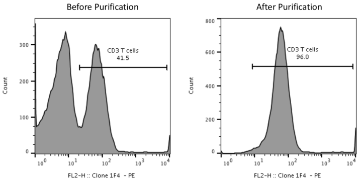
Figure 1. Purity of pan-T cells after negative magnetic isolation from PBMCs. In this representation, SD rat PBMCs were incubated with antibodies against B cells, NK cells, dendritic cells, monocytes, granulocytes, and erythrocytes and subjected to negative magnetic isolation to obtain pan-T cells by negative selection. A small aliquot was taken after selection to evaluate post-sort purity (right). Percent of CD3+ cells in PBMCs is also shown (left).
Cryopreservation and storage
Purified SD rat CD3 pan T cells were cryopreserved carefully using iQ Biosciences’ cryopreservation protocol that ensures high viability after thawing.
Cells should be stored at < -120°C once they are received, such as within a liquid nitrogen tank (vapor phase).
Additional information
| Available Size(s) | |
|---|---|
| Cell Type | |
| Format | |
| Species | |
| Tissue Type | |
| Viability | > 70% |
Tailored Experiments
Your research is important to us. Our scientists will work through each step of the process with you, including assay design, data analysis, and recommendations for future studies.
Streamlined Process
Simplify your workflow. Bypass the middle-man: at iQ Biosciences, you'll get immediate access to our biospecimen inventory, saving you both valuable time and money.
Expertise
We're experts- so you don't have to be. Augmenting years of experience in immunology and working with immune assays, our scientists stay current with the latest publications and technology.
Exceptional Service
We're here to help. We know the challenges you're facing: whether it be through expedited service or our complimentary consulting services, our team is dedicated to helping you reach your goals.
For US customers, we ship via FedEx Overnight Shipping. Shipping charges will vary per shipping address (based on ZIP code) and are estimated to be $140.
For international (non-US) customers, we work closely with you and our couriers to ensure all necessary documentation is in place for international shipments to significantly reduce the chance of delays at Customs. For the export of non-human primate samples, this includes preparing CITES permits, as well as any other documentation as required by country. Please submit an inquiry to orders@iqbiosciences.com for your estimated time of delivery and shipping charges.
Austria ![]()
Hölzel Diagnostika Handels GmbH
Tel: +49 221 126 02 66
Email: info@hoelzel.de
Web: https://www.hoelzel-biotech.com/
Canada ![]()
Cedarlane
Tel: +1 (289) 288-0001
Toll Free (North America): +1 (800) 268-5058
Fax: +1 (289) 288-0020
Email: sales@cedarlanelabs.com
Web: https://www.cedarlanelabs.com
China ![]()
BIOHUB INTERNATIONAL TRADE CO., LTD.
上海起发实验试剂有限公司
Address: Chuansha Rd #6619, Pudong, Shanghai, Zipcode: 201200 P.R.China
Tel: 0086-021-50724187
Phone: +86-15921799099
Fax: 0086-021-50724961
Email: sale3@78bio.com
Web: www.qfbio.com
European Union ![]()
Caltag Medsystems Ltd.
Email: office@caltagmedsystems.co.uk
Web: https://www.caltagmedsystems.co.uk
tebu-bio
Web: https://www.tebu-bio.com
Or Find a local contact
Germany ![]()
Hölzel Diagnostika Handels GmbH
Tel: +49 221 126 02 66
Email: info@hoelzel.de
Web: https://www.hoelzel-biotech.com/
Zageno
Web: https://zageno.com/
Ireland ![]()
2B Scientific Ltd
Tel: +44(0) 1869 238033
Fax: +44(0) 1869 238034
Email: sales@2BScientific.com
Web: https://www.2bscientific.com
India ![]()
Cell & Gene BioSolutions Pvt. Ltd.
#478 C, SLV Complex, Raghavendra Swamy Mutt Road
Opp. Turahalli Water Tank, Turahalli, Subramanyapura Post
Uttarahalli Hobli, Bengaluru-560061, Karnataka, India
Phone: +91 97317 14670
Phone: +91 98809 25033
Email: info@cgbios.com
Web: www.cgbios.com
Japan ![]()
Cosmo Bio Co., Ltd.
Tel: +81 (03) 5632 9610
Fax: +81 (03) 5632 9619
Email: nsmail@cosmobio.co.jp
Web: https://www.cosmobio.co.jp
Qatar ![]()
Sedeer Medical Services and Trading LLC
Tel: +974 4434 9191
Email: info@sedeer.com
Web: https://sedeer.com/
Singapore ![]()
Omnicell Pte Ltd
Tel: +65 6747 0201
Email: enquiry@omnicell.com.sg
Web: https://omnicell.com.sg/</a
South Korea ![]()
BioClone
Tel: +82-2-2690-0058
Email: bioclone@bioclone.co.kr
Web: http://www.bioclone.co.kr
Switzerland ![]()
Hölzel Diagnostika Handels GmbH
Tel: +49 221 126 02 66
Email: info@hoelzel.de
Web: https://www.hoelzel-biotech.com/
Taiwan ![]()
Hycell International Co. Ltd.
Tel: +886-2-2877-1122
Fax: +886-2-2876-1520
Web: http://www.hycell.com.tw
United Kingdom ![]()
2B Scientific Ltd
Tel: +44(0) 1869 238033
Fax: +44(0) 1869 238034
Email: sales@2BScientific.com
Web: https://www.2bscientific.com
Caltag Medsystems Ltd.
Tel: +44 (0)1280 827460
Fax: +44 (0)1280 827466
Email: office@caltagmedsystems.co.uk
Web: https://www.caltagmedsystems.co.uk
tebu-bio
Tel: +44 (0)1733 421880
Fax: +44 (0)1733 421882
Email: uk@tebu-bio.com
Web: https://www.tebu-bio.com
Zageno
Web: https://zageno.com/
United States ![]()
Fisher Scientific
Tel: 1-800-766-7000
Web: https://www.fishersci.com
Quartzy
Web: https://www.quartzy.com
VWR International
Tel: 1-800-932-5000
Web: https://www.vwr.com
Zageno
Web: https://zageno.com/